The human heart is located in the chest cavity in the middle of the lungs and behind the sternum (slightly to the left of the sternum or “breastbone”). It is roughly the size of a fist and one of the hardest working organs in the human body. The heart is responsible for pumping blood to all extremities after it is oxygenated. The blood is oxygenated by the lungs requiring the heart to interact in close proximity to it.
By providing this blood to various parts of the body, those parts receive the oxygen and other vital nutrients that they need in order to do their various tasks. They pump the blood through vessels known as arteries (carrying the blood from the heart) and veins (which return the blood to the heart). The heart typically moves 2,000 gallons of blood daily.
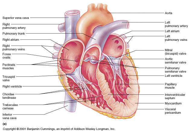
The heart consists of four chambers serving distinct functions: The upper chambers are called the left and right atria, and the lower chambers are called the left and right ventricles. A layer of muscle called the septum separates the left and right atria and the left and right ventricles. The left ventricle is the largest and strongest chamber in the heart. The left ventricle’s chamber walls are only about a half-inch thick, but they have enough force to push blood through the aortic valve and into the body.
Valves regulate the flow of blood between chambers of the heart. Logically, there are also four of these: The tricuspid valve regulates blood flow between the right atrium and right ventricle. The pulmonary valve controls blood flow from the right ventricle into the pulmonary arteries, which carry blood to the lungs to pick up oxygen. The mitral valve lets oxygen-rich blood from the lungs pass from the left atrium into the left ventricle. The aortic valve opens the way for oxygen-rich blood to pass from the left ventricle into the aorta, the body’s largest artery, where it is delivered to the rest of the body.
There is a complex and highly essential system of regulation that allows these chambers to contract and expand in the necessary rhythm for it them to provide their vital function. Electrical impulses from the heart muscle (the myocardium) cause the heart to contract. This electrical signal begins in the sinoatrial (SA) node, located at the top of the right atrium. The SA node is usually referred to in layman’s terms as “the pacemaker.” An electrical impulse from this natural pacemaker travels through the muscle fibers of the atria and ventricles, causing them to contract. Although the SA node sends electrical impulses at a certain rate, the heart rate may still change depending on various environmental factors such as physical demands, stress, or hormonal factors.
Surrounding the heart is the pericardium, a fibrous double-walled sac. It is a very tough membrane that contains the heart and the beginnings of major vessels and nerves. It is filled with a fluid that protects the heart from external jolts and pressure.